This hero is built with a flex layout, aligned and justified so that the content will always be centered horizontally and vertically. To change this section’s background, select the “Hero Overlay” then scroll to the background section of the Style panel and replace the image. You can also adjust the opacity of the overlay’s black background for better contrast.
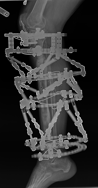
Patient walking with a stick. Main problem is still knee pain. Unfortunately this was expected from the beginning. No major pin site problems. Clinically everything looks well and progressing toward the union. Bone regenerate is also maturing satisfactorily.
From the regenerate perspective we can remove the frame, but not completely convinced that the proximal fracture has united.

Plan:


